


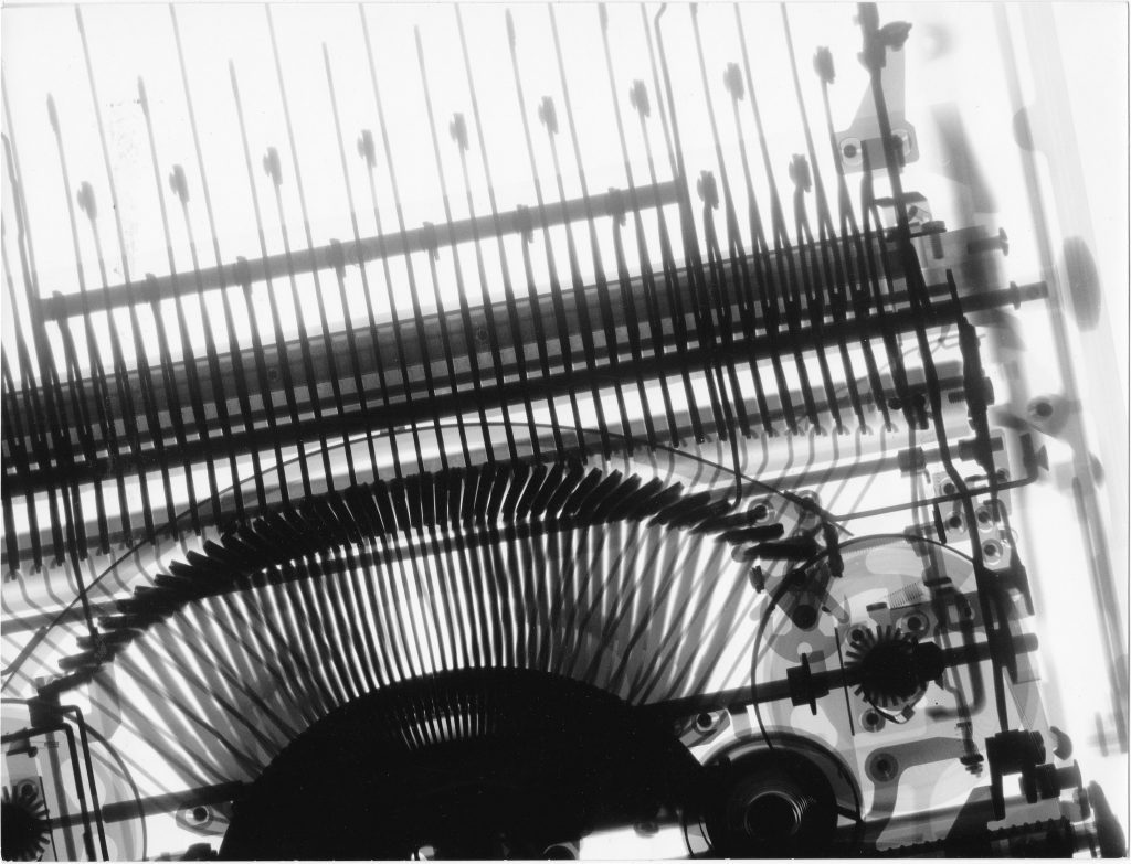

X-Ray Series Hard Radiation (1954-1958)
H. Franke first became interested in artistic experimentation while working on his dissertation, which focused on electron optics. The aesthetic quality of electron microscope images inspired him to explore the possibility of using scientific photography instruments for experiments solely intended to create aesthetically interesting images, rather than for research. Early on, he also considered converting this type of laboratory equipment into art machines. The first group of works to emerge from this concept used X-ray equipment.
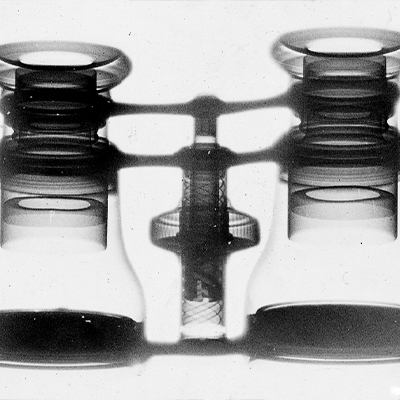
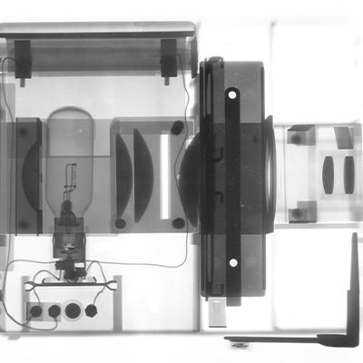
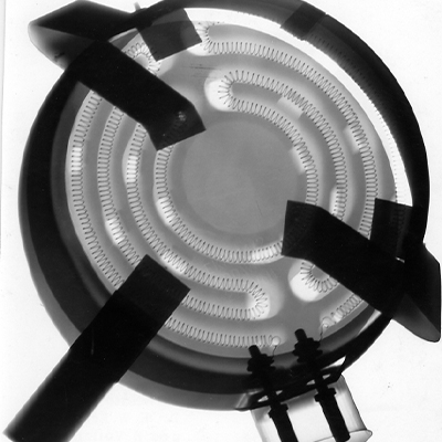
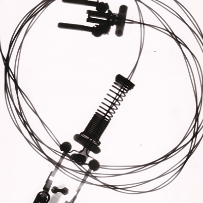
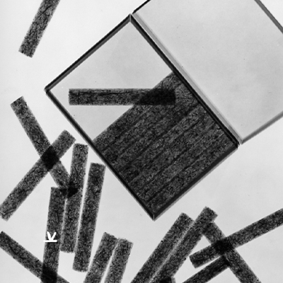
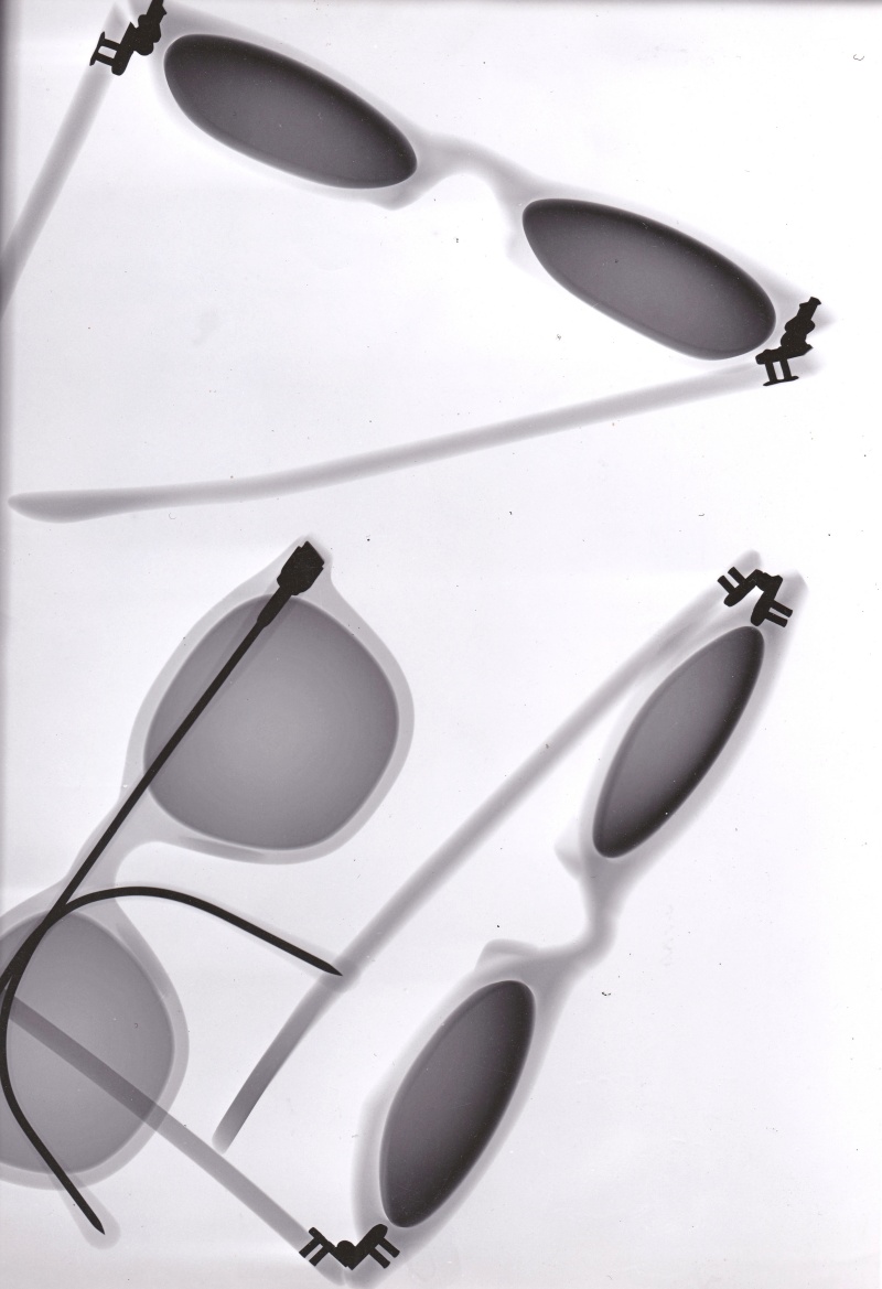
His work in medical technology at Siemens Erlangen in the early 1950s enabled him to conduct experiments in various areas of the electromagnetic spectrum. Franke gained access to this equipment through colleagues in Siemens’ research and development departments, who helped with the technical implementation of his work. Franke used hard X-rays, similar to those used for material inspection. These X-rays can penetrate materials such as stone or metal. In this series, however, Franke examined ordinary objects, such as cigarette paraphernalia, optical instruments, and items from his office and the kitchen.
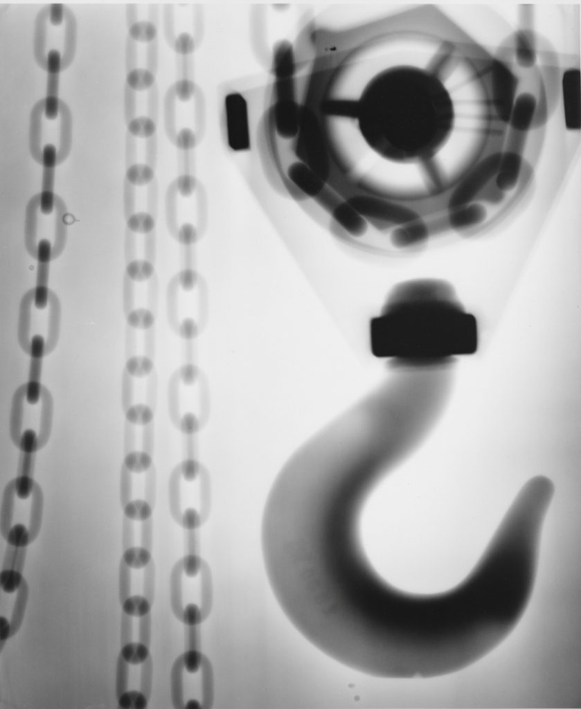
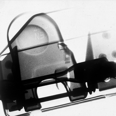
Franke was also allowed to use a very special piece of medical equipment in the development labs on one occasion. The Siemens Betatron, which was a milestone in medical radiation therapy in the 1950s, was the first large-scale, technically advanced device for cancer treatment. It is a circular electron accelerator that accelerates electrons to very high energies. The electrons could be used directly to irradiate superficial tumors, or they could be converted into high-energy X-rays (bremsstrahlung) using a converter plate for deep-seated tumors. This version was used for this X-Ray.